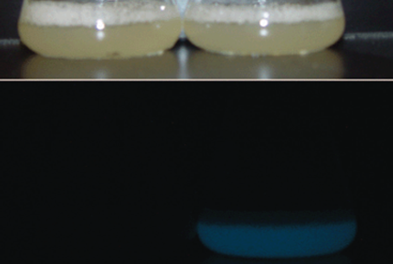
Lac operon: Two gems from the early days (1|2)
by Christoph
When I recently wrote about noise in the basement of the lac operon, I stumbled across two papers in said basement, both of which I – and not only I – consider gems of early molecular biology: Isolation of the lac repressor (Gilbert & Müller-Hill, 1966), and The nucleotide sequence of the lac operator (Gilbert & Maxam, 1973). Disclosure: Both papers were subjects of my graduation exam (biochemistry) in 1984, which probably explains my long-standing fondness for them. Here in part 1, I will dust off the lac repressor story a bit to make this gem sparkle again...
To give you an idea of the vibes in which the hunt for the lac repressor began 65 years ago, I quote below from the PaJaMo paper by Pardee, Jacob & Monod (1959), which Jon Beckwith said was "a flash of clarity in the murky sea of vaguely outlined theories that had been proposed up to this time" for genetic regulation. You've been warned, but if you're curious to navigate this 'murky sea...,' I recommend as compass Who discovered messenger RNA? by Matthew Cobb (2015) (Open Access). Now the PaJaMo quote:
(5) … Although the wide occurrence of repression effects is certain, the situation revealed with the present system, namely a genetic "complex" comprising, besides the "structural" genes (z, y) a repressor-making gene (i) whose function is to block or regulate the expression of the neighboring genes is, so far, unique for enzyme systems.
...
(6) Implicit in the repressor model are two critical questions, ... These questions are:
(a) What is the chemical nature of the repressor? Should it be considered a primary or a secondary product of the gene?
(b) Does the repressor act at the level of the gene itself, or at the level of the cytoplasmic gene-product (enzyme-forming system)?
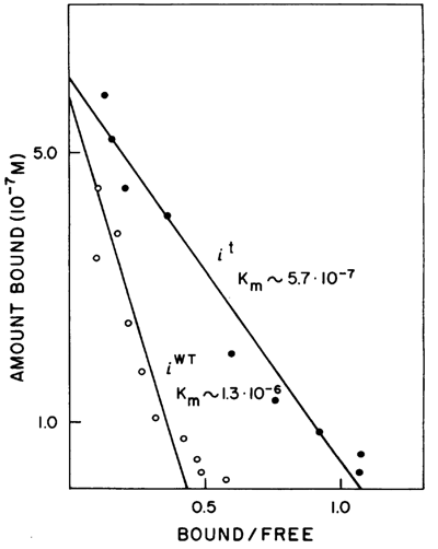
FIG. 4. The binding constants of the wild-type and i t mutant strains. Samples isolated from isogenic wild-type and i t diploid strains were dialyzed against many different concentrations of 14C IPTG as described under Fig. 2. The amount of IPTG bound at each external IPTG concentration, normalized to a protein concentration of 10 mg/ml, is plotted against the ratio of bound to free IPTG. Single-side binding should obey the relation: (Amount bound) = (number of sites) – Km (bound/free). Source. Frontispiece: Model of lac repressor tetramer bound to two lacO operators, looping-out of intervening DNA. Source
(Addressing the questions in reverse order) Question (6b) may seem a bit far-fetched from today's perspective – we now know that the repressor (LacI) binds to the gene itself, that is, to the operator (lacO), see part 2 – but based on their genetic experiments Pardee et al. (1959) could not rule out the possibility that the repressor binds to and blocks the 'cytoplasmic gene-products', β‑galactosidase (LacZ) and permease (LacY) (or binds to and blocks George E. Palade's microsomes (ribosomes), the 'enzyme-forming system', known since 1955). To raise the question of the chemical nature of the repressor (6a) was obvious, but it was intriguing to ask whether it was the primary or secondary gene product of the repressor gene, lacI. The 'primary gene product' would be an RNA transcript, An Unstable Intermediate Carrying Information from Genes to Ribosomes for Protein Synthesis, whose existence was first demonstrated by Brenner et al. (1961) and that we now know as messenger RNA (mRNA). The 'secondary gene product' would be the translation product of the primary transcript, a protein.
In fact, Jacob & Monod (1961) favored in their landmark paper Genetic regulatory mechanisms in the synthesis of proteins a model in which the lac repressor was the primary transcript of the lacI gene, the mRNA, which, however, could not be substantiated. Meanwhile, Gilbert & Müller-Hill had other ideas...
Isolation of the lac repressor
Gilbert & Müller-Hill felt they were on the right track with their assumption that the lac repressor is a 'secondary gene product' of the lacI gene, a protein, since they (and others) had succeeded in isolating Suppressible regulator constitutive mutants of the lactose system in Escherichia coli (that is, nonsense mutations in the lacI gene with a lacI c phenotype that can be partially suppressed by supressor tRNAs). They set about designing an assay that "detects and quantitates this repressor by measuring its binding to an inducer, as seen in this case by equilibrium dialysis against radioactive IPTG (isopropyl-thio-galactoside)." And they continue: "we desired some means that would not depend on the specific models that might be imagined for the actual mechanism of repression. The minimal assumption on which the assay is based is that there should be some interaction between the repressor and the inducer."
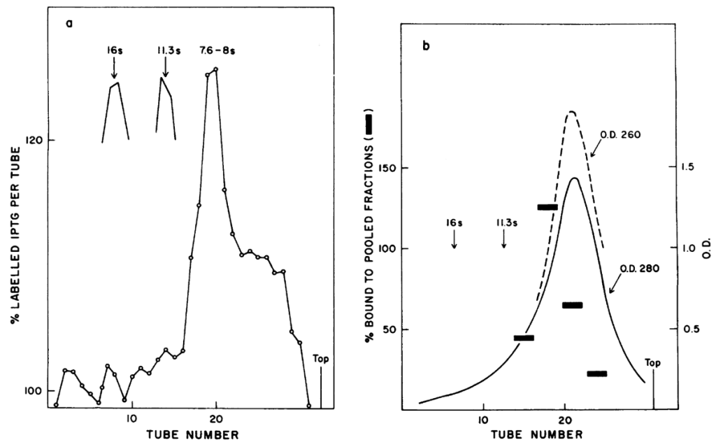
FIG. 5. Sedimentation of the repressor. Samples of 0.2 ml of a concentrated repressor fraction were layered on 5‑ml gradients, from 5 to 30% glycerol, in TMS buffer. The gradients were spun for 8.5 hr at 50,000 rpm at 40°C. In one tube, catalase and β‑galactosidase were run as sedimentation markers. For the experiment shown in (a) the repressor fraction was brought to 2.5×10⁻⁷ M 14C IPTG and layered on a gradient containing 1.2×10⁻⁷ M 14C IPTG. After the centrifugation, two-drop samples were collected directly into scintillation vials and counted with Bray's solution. The background level in the gradient is about 1000 cpm; the counts are plotted as a per cent of the background level (open circles). For the experiment shown in (b), a parallel gradient was run without IPTG. The optical density was measured on each two-drop sample at 260 mμ (=nm) (—-) and 280 mμ (=nm) (——). Three-tube samples were pooled and placed in dialysis sacs. Their volumes were reduced by burying the sacs under dry G 200 Sephadlex for 1 hr. Then the samples were dialyzed against 14C IPTG. The excess bound, corrected to 0.100 ml final volume inside the dialysis sac, is plotted as a bar. superimposed on both experimental curves is the position of the markers taken from a parallel gradient. Source
Conceptually, their approach was similar to the classical method for isolating enzymes: gradually refined fractionation of total cellular protein and in vitro testing of the individual fractions by an enzyme assay. Since they had a few clues that IPTG is not metabolized in the cells – such as lactose, which is converted to allolactose – but acts directly as an inducer, this approach seemed straightforward (14C‑labelled IPTG had just become commercially available then).
Initial attempts to tag the lac repressor by affinity labeling with IPTG failed (as Müller-Hill later recalled). In retrospect, this was actually advantageous, as it brought them to use equilibrium dialysis as a detection method for the repressor (much like an enzyme assay, which would have been impossible with affinity labeling). The problem was obtaining sufficient quantities of lac repressor! They estimated from Kinetic studies of beta-galactosidase induction that "the wildtype repressor should have an affinity of the order of 6×10⁻⁶ M for IPTG, and if any binding is to be detectable [by equilibrium dialysis], the repressor concentration must exceed a few times 10–6 M." They "would have to fractionate and concentrate the repressor by at least a factor of 100, working blindly, before detecting any effect." They overcame this hurdle in three steps: first, they isolated a lacI mutant (i t) with a ~ten-fold higher Km for IPTG (see FIG. 4.), second, they made a diploid derivative of the i t mutant strain (that is, the i t allele on a F' plasmid in addition to the chromosomal copy) to have twice the repressor concentration at the start, and third, they fractionated late‑log cell extracts with spermine and ammonium sulfate to have more lac repressor per mg protein. Thus they obtained "material that will draw the inducer into a dialysis sac to a concentration 1.5–2 times that outside thes ac (50–100% excess binding) at a protein concentration of 10 mg/ml." To this material, "The labeled IPTG is bound reversibly; it can be freely dialyzed into, out of, and even into the sac again. This is illustrated in Table 1."
Among the numerous negative and positive controls – all of which gave the expected results – one stands out as, yes... elegant: An identical parallel purification was run on 50-gm lots of diploid i t control cells and diploid (amber-suppressor-sensitive) i – cells that express a truncated lac repressor (see above). At the end of the purification, samples of each protein solution were dialyzed against several different IPTG concentrations: no binding was observed for material from the i – cells.
Gilbert & Müller-Hill determined a molecular weight of 150–200 kDa for their preparation of the lac repressor obtained by glycerol gradient centrifugation. From the yield of lac repressor isolated from the total amount of processed protein, they calculated that the cells contained no more than about ten repressor molecules per lacI gene copy. They phrase the actual Aussagekraft of their experiments carefully and list the properties of the lac repressor:
The ability to bind IPTG is not attacked by RNase or DNase. It is destroyed by pronase. The binding site can be inactivated by temperatures above 50°C. These are properties of the part of the molecule that interacts with the inducer. As yet there is no way of seeing the rest of the molecule.
This is the isolation of the lac repressor, no question, but not its purification according to the high standards of the protein specialists (see Kornberg (2009) Why purify enzymes?). The impurities in their preparation mentioned in the caption to Fig. 5. would have been apparent in Coomassie-stained SDS-PAGE, but this technique was not yet lab standard at the time (popularized by Laemmli in 1970). Above all, they would have seen in denaturing SDS-PAGE that their (native) 150–200 kDa lac repressor is in fact a tetramer, which became apparent when Beyreuther et al. (1973) determined The amino-acid sequence of lac repressor: 347 amino acids (38.5 kDa). Friedman et al. (1995) determined the Crystal Structure of lac Repressor Core Tetramer and Its Implications for DNA Looping (Figure: PDB-Model, Frontispiece). The Structural Analysis of Lac Repressor Bound to Allosteric Effectors by Daber et al. (2009) revealed that binding of IPTG induces a conformational change in the region connecting the N- and C‑terminal domains to the protein, thereby preventing efficient binding of the dimer to operator DNA (see here a diagram).
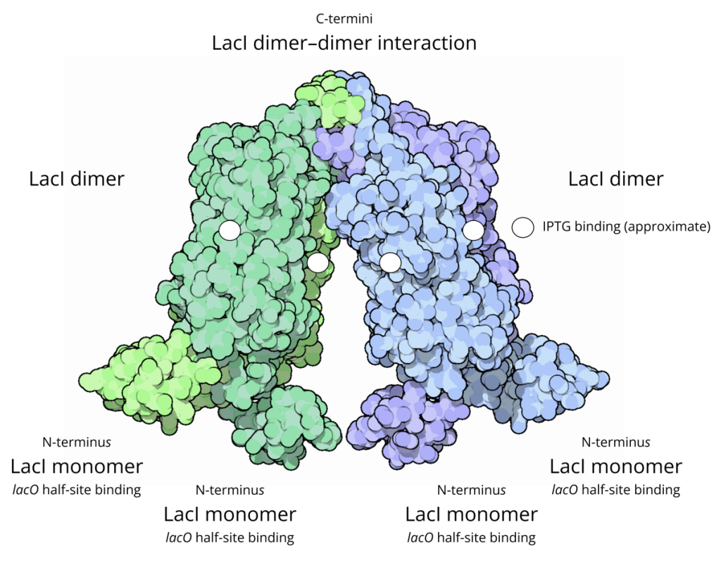
Figure: PDB‑Model of a the lac repressor tetramer (with my added descriptions)
I hope you agree that it was worth dusting off this gem. Without further ado, let me finish by quoting two sentences from an essay by Benno Müller-Hill (1990) titled, you guessed it, The Isolation of the lac Repressor: "One day [in autumn 1966], late in the afternoon, I saw Monod standing in the door. 'Hello' he said. 'Hello' I answered. And then he simply said 'Benno, after all it was pedestrian', bowed and disappeared, leaving me baffled for a long time. Now, many years later, I like to interpret his comment as 'slow, down to earth, with only a few technical crutches' and think it was, after all, a compliment."
We'd be delighted to hear from you! You can comment below, send us an email or reach us via Bluesky or Mastodon. Please subscribe to get email notifications of our new posts.