Seeing What Isn't There
Pictures Considered #6
by Elio (prompted by Christoph's suggestion)
Mapping deletion mutations by genetic tools is one thing, seeing them displayed along the length of a DNA molecule is quite another. But how can one see what isn't there? In 1968, Ronald Davis and Norman Davidson did just that—visualizing deletion mutations in the genome of coliphage λ. They reasoned that if the double-stranded DNA chromosomes of wild type phage and of a deletion mutant were dissociated and allowed to anneal together, a deletion would show up in hybrid chromosomes as an (unannealed) single-stranded loop flanked by double-stranded (annealed) DNA regions. They set to work in the lab and successfully confirmed this idea.
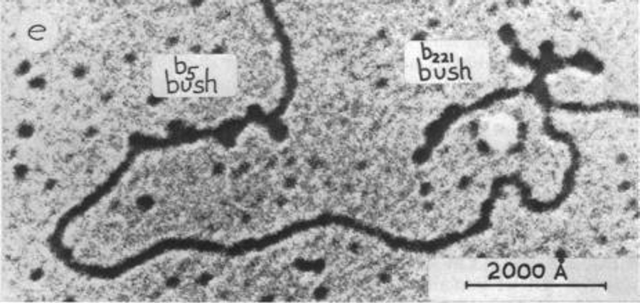
A reannealed DNA molecule showing two sites of deletion, "bushes" b5 and b211. Source
Under the electron microscope, such hybrid DNA molecules were seen to be mostly ordinary double-stranded DNA punctuated by occasional visible structures they called "bushes." Each bush was formed by the collapse of a loop of single-stranded DNA, thus likely represented the location of a deletion mutation. Although the contour length of a bush could only be estimated, their location along the DNA molecule could be accurately measured. Sure enough, the position of the bushes corresponded to the location of the deletions as estimated from recombination experiments. The fraction of the DNA deleted, as measured in the photographs, was consistent with the observed changes in the buoyant density of the wild type and mutant phages in cesium chloride density gradients—a further confirmation of their interpretation of the bushes. The authors developed a bunch of controls to ensure that they were not looking at artifacts. Thus, they could conclude that genetic measurements of distances reflected a physical reality. On a gross scale, this had been known from the position of the bands in the giant salivary gland chromosomes of Drosophila, but this study established that the gene chromosome map corresponded to a physical chromosome on a molecular level.
In case you wonder why the authors didn't simply sequence the molecules, well, practical DNA sequencing didn't become available for another five years or so. Once sequencing became a routine procedure, it obviated the necessity to come up with clever approaches to many basic questions. Ah, well...
The paper ends with the following statement: After completing this manuscript we have learned that the technique described herein has been independently conceived and developed by Westmoreland, Szybalski, and Ris, who have obtained results which are generally similar to ours. It happened even then!
Reference
Davis RW, Davidson N (1968). Electron-microscopic visualization of deletion mutations. Proc Natl Acad Sci USA, 60 (1), 243−250. PMID 5241526