Larval Settlement
by Roberto
Animals originate, evolve, and undergo development in a microbial world. This point was elegantly brought home as the "new imperative" for biology in an influential perspective by McFall-Ngai et al. in 2013 (highlighted by Elio here️). The fact that ubiquitous bacterial-animal interactions are mediated by the exchange of molecules, i.e., chemical ecology, was reviewed soon thereafter by Cantley and Clardy. A beautiful example of how animal development is guided by bacterial chemical cues is the metamorphosis of the bottom-dwelling marine worm Hydroides elegans.

Fig. 1. Adult and larval stages of Hydroides elegans. (a) Aggregations of adult H. elegans on extruded plastic mesh. Several individuals have their crowns of tentacles extended for feeding. (b) Metamorphically competent larva of H. elegans. Arrow points to apical cilia that are probably used to sense inductive cues on biofilm bacteria. Source. Frontispiece: Adult Hydroides elegans. Source
Larvae of marine invertebrates only survive and metamorphosize into juveniles (and eventually adults) when they settle on the "right spots " at the bottom of the sea. Yet they do not settle randomly. How do the larvae find these spots? Invariably, these are surfaces covered with microbial biofilms. The process of submerged surface colonization, first by bacteria and then by sedentary animals such as barnacles, is often referred to as biofouling. (As an aside, I offer my very personal opinion on this term, which I find horribly anthropocentric. What gets fouled? The invasive surfaces that humans insist on keeping pristine for economic reasons, e.g. hulls of ships. Rather than biofouling, I see beautiful ecology.)
H. elegans is a wonderful model system for the study of larval settlement. Thanks in large part to the work carried out by Michael Hadfield and colleagues in Hawaii since 1990, the life cycle of this worm, particularly the settlement on bacterial biofilm, is now understood in detail. They were able to cultivate the animal in the lab and thus manipulate its environment. In the process they were able to isolate and characterize strains of bacteria that were particularly good at inducing larval settlement. Among these were isolates of Pseudoalteromonas luteoviolacea.

Fig. 2. Top: Electron micrographs of several functional states of the contractile structures. Scalebar = 100 nm. Bottom: Schematic of the functional states. For full legend of figure see source.
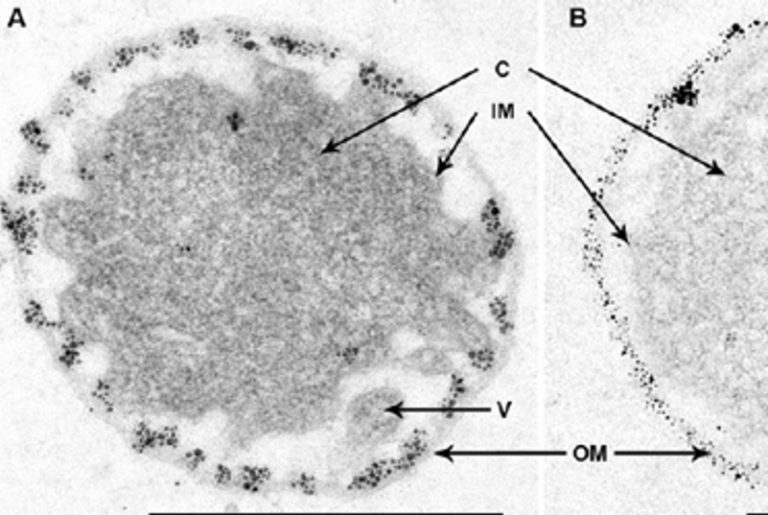
The power of genetics led to the discovery of a mechanism through which P. luteoviolacea signals the larva to settle on top of the biofilm and initiate development. First came the isolation of bacterial mutants that no longer recruited the larvae to the biofilm and thus failed to metamorphosize. Sequence analyses of the interrupted genes indicated that their products were similar to tailocins, phage tail-like bacteriocins. This insight led to microscopic analyses showing that contractile structures of the bacteria were indeed involved in larval settlement and metamorphosis (Fig. 2). Subsequent results identified a protein (metamorphosis inducing factor 1 or Mif1) that was injected into the larvae through the contractile structure and which initiated the developmental transformation by increasing diacylglycerol, thus triggering a phosphotransferase signaling cascade. This set of beautiful results might lead one to believe that there is a universal molecular mechanism through which larvae from marine invertebrates can identify good spots for settlement and metamorphosis. Except... there's more than one way to skim a biofilm.
In the initial search for bacterial strains that induced larval settlement and metamorphosis, Cellulophaga lytica also came up. Yet, its genome does not encode any phage tail-like structures. The hunt was on for another molecule responsible for the inducing activity. Through a rigorous (and laborious) activity-guided fractionation, the authors of a recent study purified the inducing molecule. It was the lipopolysaccharide (LPS) of C. lytica's outer membrane.

Fig. 3. The general structure of the lipopolysaccharides (LPS) of Gram (-) bacteria. LPS is the main component of the outer layer of the outer membrane. Adapted from source.
For the uninitiated, the LPS is the unusual lipid that makes up the outer leaflet of the outer membrane of Gram-negative bacteria. LPS can be divided into three components. Lipid A is highly immunogenic and is recognized through the TLR4 receptor of the innate immune system. Lipid A is connected to the core oligosaccharide which in turn is connected to a highly variable polysaccharide known as the O‑side chain. Its variability is both in the composition and number of the repeating oligosaccharide units. When Gram-negative bacteria infect a mammalian host, antibodies are often generated against the O‑side chain. Thus its other name, the O‑antigen. The variability in O‑antigen structure can form the basis for serotyping of bacterial strains (e.g., the pathogenic E. coli O157). The part of the LPS from C. lytica that induces H. elegans to settle and metamorphosize turned out to be the O‑antigen. Not surprisingly, not all C. lytica strains induce development because O‑antigens are highly variable. I find this high degree of selectivity by the larvae fascinating and, to me at least, indicative of coordination between bacteria and animal that is more than just the recognition of a good spot to settle on. Are there any other steps in the life of the animal that are guided by its association with biofilm bacteria? Has there been some co-evolution of bacteria and animal? We'll just have to wait and see. I think there's great potential for discovery in studying this fascinating animal-bacterial interaction.
Do you want to comment on this post? We would be happy about it! Please send us an email, or comment on Mastodon or Bluesky.