Getting to grips with the intangible
by Christoph
Imagine the E. coli nucleoid as a swig but of a highly viscous tangle of a 2 nm thin and ~1.5 mm long DNA polymer, decorated with and highly compacted by a plethora of proteins, and somehow suspended in the cytoplasm of a cell with a volume of ~1.3 µm3. It's virtually impossible to "grip" the nucleoid, to measure physical dimensions like shape and volume of this membrane-less organelle, or compartment, in bacterial cells that contains (most of) their genetic material. Just the other day, I stumbled across the pre‑print, a not yet peer-reviewed, valid publication of a study that really sent me into (scientific) highs because of the experimental approach to measuring physical properties of the nucleoid in live cells, an approach that has, to my knowledge, not been pursued consequentially so far. And, without overstating it, this approach is comparable to supplementing conventional medical diagnostics of X‑ray and ultrasound examinations with minimally invasive endoscopy. Yingjie Xiang and colleagues from Christine Jacobs-Wagner's lab at Stanford University, USA, addressed the "...somehow suspended" from my above sentence. Here, I will deal with their calculation of the average "mesh size" of the nucleoid tangle from measurements of live cells.

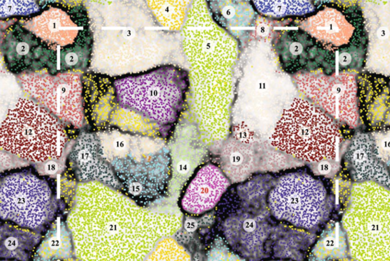
Figure 1. Estimation of the average DNA concentration in the nucleoid region in B‑period E. coli cells. A Fluorescence images of representative DAPI-stained CJW6324 cells in different cell cycle periods: B, C and D periods (i.e., before, during and after the DNA replication). The green and yellow outlines are the cell and nucleoid contours detected by Oufti. B Plot showing the relative area of the SeqA-mCherry signal as a function of cell area. The contour lines (from light to dark color) represent the 25%, 50% and 75% probability envelopes of the data. C Probability density function of the estimated nucleoid volume in B‑period cells. D Probability density function of the average DNA concentration calculated based on the total mass of the E. coli chromosome and the nucleoid volume measurements shown in panel C. Frontispiece: The ribosome tomogram (from Fig. 6D) was obtained after Gaussian smoothing (𝜎 = 1.2 pixel, 21 nm) of the top map to more clearly show subcellular regions with high and low density in ribosomes. Probability density (×106): 1.6 dark red − 0.6 dark blue. Source
Note. STC has featured the bacterial nucleoid repeatedly: in a post from 2008 by Conrad Woldringh, in 2013, in 2014 by Suckjoon Jun, in 2019 by Roberto jointly with, again, Conrad Woldringh, in May this year by Elio, and just recently by Roberto who delved deeper into the intriguing story of the interaction of the Dps protein with the E. coli chromosome.
Xiang et al. had to find conditions where the cells have exactly one chromosome in order to calculate the nucleoid volume and the DNA concentration within. From this, they derived its average mesh size. It is known since the fundamental study of Cooper and Helmstetter (1968) that E. coli B/r cells have discrete B‑, C‑, and D‑periods when grown in nutrient-poor medium at 37°C, and, importantly, B‑period cells have exactly one non-replicating chromosome (see here for a diagram.) Here, the authors identified B‑period cells within a population of E. coli K‑121) cells grown at 37°C in glycerol-supplemented M9 minimal medium by examining cell size and spatial pattern of a DNA replication marker, SeqA‑mCherry (Figure 1A+B). Some background here: the SeqA-mCherry fusion protein binds newly synthesized hemi-methylated DNA resulting in fluorescent foci in cells actively replicating DNA (C‑period). In contrast, SeqA-mCherry is diffusely distributed in cells before (B‑period) and after DNA replication, shortly before cell division (D‑period).

Figure 2. Estimation of the average nucleoid mesh size. A Schematic of an assembled dodecahedron-shaped nanocage-GFP particle (⌀ 25 nm). B Fluorescence images of nanocage-GFP particles assembled in E. coli cells (CJW6340). C Example of nanocage-GFP particle trajectory. The gray outline indicates the cell contour detected by the Oufti software using the corresponding phase image of the cell. E Probability density map of the localization of nanocage-GFP particles. The density map was constructed by plotting the normalized 2D histogram of the relative particle positions. F Probability density maps of the localization of GFP-μNS particles as a function of their size. The positions of the particles (n = 133,692) were determined relative to the cell contours. Particles were then binned by their sizes, as indicated. For each particle-size bin, a probability density map of particle localizations was constructed, as described in panel E. Source
The authors selected 19,510 B‑period cells, measured the contour of their DAPI-stained nucleoids (using appropriate software, of course) and calculated the average nucleoid volume as ~0.7 μm3. Although DAPI staining does not easily distinguish between denser and less-dense nucleoid regions known from earlier electron microscopy and more recent super-resolution microscopy studies, Xiang et al. found a surprisingly narrow distribution of nucleoid volumes (Figure 1C). With the known genome size of their E. coli K‑12 strain (4.6 Mb) they calculated a DNA concentration in the nucleoids of 7.1±1.0 mg/ml (Figure 1D). (Calculating with a molecular mass of 618 g/mol per base pair, I arrive at a DNA concentration of 7.3 mg/ml for the nucleoid, which is an acceptable deviation.)
Assuming that the cytoplasm is an ideal solvent for the nucleoid, and using the measured values for its volume and the DNA concentration within, they obtained a value for the mesh size of 22 nm in a complex formula describing the physico-chemical properties of a semi-dilute polymer in a solvent (I won't go into the details but aficionadas y aficionados can appreciate the formula here.) If this were the case, the diffusion by simple Brownian motion of cytoplasmic particles larger than 22 nm should be impeded by the nucleoid. Spoiler: that's not the case (ergo the cytoplasm is not an ideal solvent for the nucleoid.) Can something as abstract as "mesh size" actually be measured in vivo?
Xiang et al. expressed the GFP-tagged, artificial protein I3-01 in E. coli that self-assembles into a 'nanocage' of 25 nm in diameter and is unlikely to form specific interactions with the nucleoid or other cytoplasmic components (Figure 2A). Basal, that is, leaky expression of this GFP-tagged protein in E. coli B2) was sufficient to produce a single fluorescent nanocage per cell in a fraction of the population (Figure 2B). Single-particle tracking experiments revealed no evidence of steric hindrance by the nucleoid as the nanocage appeared to diffuse unrestrained and at constant "speed" throughout the cytoplasm (Figure 2C). Lastly, a calculation of the relative positions of 2,500 nanocages inside cells (normalized because of varying cell lengths) showed that the probability density of their localization is uniform throughout the cytoplasm (Figure 2E). To repeat the above: the free diffusion of a particle with a diameter of 25 nm throughout the cytoplasm is not negatively affected by the nucleoid − but what about larger particles?

Figure 3. Spatial distribution of ribosomes in E. coli tomograms. Wild-type E. coli cells (MG1655) were harvested in exponential phase, concentrated by centrifugation, deposited on the grid + vitrified in liquid ethane. Using cryo-FIB milling, a thin lamella sample of 155–260 nm thickness was obtained for the cryo-ET imaging. B Example section of the tomogram (see here). D Localized ribosome structures (yellow) with the positions and orientations determined in the tomogram shown in panel B. Source
Xiang et al. obtained particles with diameters ranging from ~50 to ~200 nm by expressing GFP-μNS, a GFP‑tagged reovirus protein, from the chromosome of E. coli CJW46173) following IPTG induction for up to 120 min. GFP-μNS assembles into globular fluorescent particles, usually one per cell, and is unlikely to display a significant affinity to components of the E. coli cytoplasm. They tracked 133,692(!) GFP-μNS particles and calculated their relative positions in the cells and then binned all GFP-μNS particles by size. For each size bin, they constructed a probability density map of the relative particle position in the cells (Figure 2F). In previous experiment, they had already calibrated the GFP-μNS particles for their size according to the corresponding fluorescence intensities. Note that the growing cells had, of course, different sizes (B‑, C‑, and D‑period cells.) A midcell constriction largely devoid of GFP-μNS particles becomes apparent with increasing GFP-μNS particle size by the size-normalization procedure that also included the pre-division cells. For particles in the bin with the smallest average particle size (⌀ 51 nm, Figure 2F leftmost), the probability density map appeared indistinguishable from that obtained for nanocage-GFP particles (Figure 2E), indicating that these particles are not excluded by the nucleoid. However, this pattern changed with increasing particle size, as particles from size of 58 nm on gradually display an increased probability for localization at the cell poles, away from the nucleoid (Figure 2F). The largest particles (⌀ 150 nm) were almost completely excluded from the nucleoid, as shown by their high density at the cell poles and the near-zero density elsewhere in the cell. Because there is no apparent threshold size, these ensemble results cannot provide an exact value. Xiang et al. give the apparent average nucleoid mesh size as ~50 nm, therefore.

Figure 4. Pixel intensity correlation between DNA and ribosome fluorescence signals. A Fluorescence micrographs showing an example cell (E. coli CJW7020) with a strong negative correlation (Spearman's 𝜌 = –0.89) between the DNA and ribosome fluorescence signals. The cell outlines are depicted as white solid lines, whereas the nucleoid outlines are shown as either black or white dash lines. The images are shown using either the conventional gray scale (top row) or a color scale (middle row). The bottom row shows only the pixels enclosed well within the nucleoid, at least two pixels away from the nucleoid outline. These pixels were used for the correlation calculation. B Similar to panel A, except for showing an example cell with a more common negative correlation (Spearman's 𝜌 = –0.60) between DNA and ribosome fluorescence signals. C Scatter plot showing the pixel intensity correlation between DAPI and L1-msfGFP signals obtained in the cell shown in panel A. D Similar to C, except for showing the corresponding pixel intensity correlation in the cell shown in panel B. E Probability density function of pixel intensity correlations calculated among the cell population (n= 1,126). Source
If larger particles (⌀ >50 nm) cannot diffuse freely throughout the nucleoid in contrast to smaller particles (⌀ 25 nm), what would it look like if thousands of such smaller particles, singly or in chains, were used to probe the nucleoid? For example in a cryo‑electron microscopic "snapshot" of a cell, a technique that is less prone to artifacts than conventional electron microscopy. It's long known among electron microscopists that 70S ribosomes are particularly well suited targets because they are electron-dense, uniform in size (⌀ ~21 nm), present in high numbers in cells (7×10³ − 7×10⁴ per E. coli cell depending on the growth rate), and localize preferentially to cytoplasmic space depleted for the nucleoid (maybe you recall this cryo-ET image of an ultra-small bacterial cell from an earlier STC post.) Most, but not all ribosomes in the cells are poly-ribosomes, that is, they are arranged like beads on a mRNA "string" during translation, which would exclude them from nucleoid-denser space due to their size.4) Xiang et al. prepared E. coli MG1655 cells exponentially growing in rich medium5) at 37°C to boost their ribosome numbers − for cryo-ET and scanned lamellae, that is, ~150−250 nm-thin slices obtained by cryo-FIB milling. Figure 3 shows a longitudinal cross section of an E. coli MG1655 cell (~2x1 µm) for which 5,028 ribosomes in the analyzed ~200 nm-thin lamella were localized by a structure‑finding program that was trained on the precisely known silhouette of 70S ribosomes (this video gives a fairly good 3D impression.) You certainly see in the picture the "void" between the not completely evenly distributed ribosomes, that is, the space presumably occupied by denser portions of the nucleoid. It is thus similar to a casting mold, but one with blurred edges. This "negative imaging" of the nucleoid is what I had in mind when I titled this post: Getting to grips with the intangible. Note, however, that this is a reconstruction of the "void" in one individual cell at a single point in time. In other cells, the distribution of the particularly dense regions of the nucleoid will vary.
Xiang et al. wanted to confirm that the "void" in their ribosome tomograms was indeed filled with nucleoid material. To this end, they used E. coli CJW7020 cells producing 50S ribosomal protein L1 tagged with superfolder GFP (green fluorescent protein, Emax 525 nm) that were stained with DAPI (Emax 461 nm) for the chromosomal DNA (Figure 4). They identified the nucleoid outlines in 1,126 cells based on the DAPI signal (dotted lines in Figures 4A+B for two cells). For the calculation of the correlation between the DNA and ribosome fluorescence signals, they considered only image pixels well within the nucleoid region (Figure 4A+B, bottom row.) Doing so avoids any bias in the correlation analysis due to the known enrichment of poly-ribosomes outside the nucleoid region. With these precautions, they calculated the correlation coefficient (Spearman's 𝜌) between the fluorescence signals of DAPI (DNA) and L1-msfGFP (ribosome) at the singlexel resolution and found that even well within the nucleoid, the ribosome fluorescence signal correlates negatively with the DNA signal (two cell examples Figure 4C+D, population level n = 1,126 cells, Figure 4E). They could thus safely conclude that the "void" in their ribosome tomograms was indeed filled with nucleoid.

Figure 5. E. coli B infected by T2 phage, fixed 30 min after infection under the standard conditions, and embedded in vestopal W. Magnification 50,000×. Mature virus (v) are clearly visible. Their polyhedral heads are well preserved. The pool (po) is composed of thin filaments, but it is more dilute than that of chloramphenicol-treated cells. The dense bodies appearing in the cytoplasm are probably the chondrioids (ch) [= mesosomes, now considered EM artifacts]. Figs. 5a − 5c differ only in the washing bath between the same standard fixation and the dehydration: (a) is treated 1 h in buffer containing 0.1 M Ca++, (b) is treated for 2 h in a 1% solution of La(NO3)3 in buffer, © is treated for 2 hours in a 0.5% solution of uranyl-acetate in buffer (pH 5). All these micrographs were photographically treated in the same way, until the background appeared of the same gray shade. Fig. 5d is the same as Fig. 5a, but enlarged on a photographic paper 2 grades harder. These four pictures show that the lanthanum and especially the uranyl ions increase the contrast not only of the phages, but also of the bacterial cytoplasm, since photographic augmentation of contrast for a bacterium not treated with uranyl-acetate gives an image very similar to that of a treated cell. See Fig. 5 c+d here. Source
I wonder if Xiang et al. came up with the idea of measuring the mesh size of the nucleoid inside living cells after having seen Figure 5. Taken from a paper by Kellenberger et al., these electron micrographs show bacteriophage T2 heads assembling in E. coli B host cells ~30 min after infection (this paper was published ~60 years ago, in 1958!) The phage heads have assembled in groups − or as array, see Fig. 5c − at the edges of the "pool" (po) but not within. This "pool" is replicating phage DNA that appears structurally indistinguishable from the nucleoid in electron micrographs. The nucleoid, however, isn't there anymore as the host chromosome is degraded within minutes after infection.6) Since T2 phages have a burst size of ~120/cell and a genome size of 164 kb they "fill" the cell upon replication with an amount of long fibrillar DNA molecules that exceeds that of host chromosomal DNA by a factor of four.7) No wonder that the "pools" seen in Figure 5 could easily be mistaken as the cells' nucleoids! Like T4 phages, T2 phages have slightly elongated heads and Kellenberger et al. determined their dimensions as 32.3×71.0 nm. This is roughly 20% smaller than values you find for phage heads prepared by different methods and that vary wildly between 65×90 nm (freeze-dried), 85×98 nm (agar filtration method), and 80×114 nm. The authors assume that the reason for the "constriction" (=shrinking) of the phage heads in their electron microscope studies is the embedding procedure and dehydration. In any case, these phage heads are bona fide larger than the nucleoid's average mesh size of ~50 nm as determined by Xiang et al., and it thus makes sense to find them at the edges of the nucleoid-like phage DNA "pool" and not within (Figure 5).
Enthralling studies usually solve some puzzles, answer some questions but create just as many new ones, or make completely new questions possible in the first place. For example, it is long known that in E. coli various so-called nucleoid-associated protein (NAPs) contribute to chromosome compaction. Spurio et al. (1992) have shown for one of them, H‑NS, that overexpression is lethal and results in super-compacted nucleoids (see here). Do such nucleoids have aberrant mesh sizes? And what about the nucleoid mesh sizes of E. coli hns‑2 mutant cells that lack H‑NS but are viable, pretty healthy in fact? And then there is the long-standing observation that the intracellular concentrations of all NAPs vary considerably over the growth phases... Let experimentation begin!
Footnotes
1) Strain CJW6324 (MG1655 seqA::seqA-mcherry ftsZ::ftsZ774‑venussw).
2) Strain CJW6340 (BL21 Star (DE3) pET29b+ I3-01-sfgfp)
3) Strain CJW4617 (MG1655 ΔlacZYA::gfp-μNS) produces GFP-μNS particles of tunable sizes in response to IPTG induction.
4) Single-stranded RNA is not visible in electron microscopy unless metal-shaded during sample preparation, see Pictures Considered #1.
5) They used M9 medium supplemented with 0.2% glycerol, 0.1% casamino acids, and 1 μg/ml thiamine, which is not usually considered as 'rich medium' but in fact comes close.
6) An aside: generalized transduction with 'T even' phages is possible but done with specific mutants with defects in host-DNA degrading activity.
7) Note that it is not possible to determine the volume of the "pool" exactly as the number of replicated phage genomes is variable between cells, and the fraction of genomes already packed in the heads is unknown.