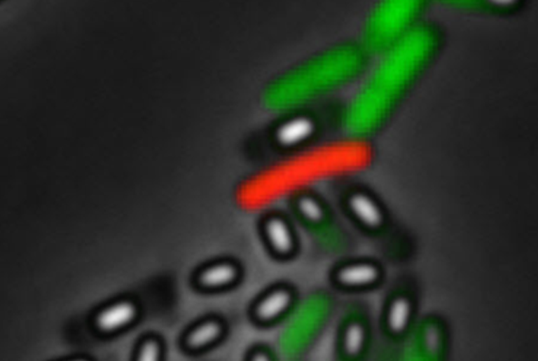
Teachers Corner: Who bears the load?
by Roberto
First, let me comment on the title I wrote. It should have read, what bears the load? Better yet, what part of the bacterial cell envelope is the load-bearing component? Whenever I taught bacterial cell biology I always started by describing the components of cell envelope, cytoplasmic membrane, cell wall and, in the Gram-negatives, the outer membrane. Then I would invariably pronounce that the cell wall, with its remarkable mesh of covalently linked glycan strands and peptide crosslinks, was the rigid load-bearing envelope component. Just looking at the structure suggested it was a stiff molecule able to bear the mechanical stresses that a bacterium might encounter in its changing environments. Right? Well, not quite… Apparently, for Gram-negatives, the outer membrane is a very important load-bearing component of the cell envelope. That is the finding that was reported in 2018 in a paper whose first author is Enrique Rojas, who worked with Julie Theriot and K.C. Huang at Stanford and is now in the faculty at NYU. The paper presents the results of a collaboration that involved not only those labs but also groups at UCSF and Madison. Overall, I found the results quite compelling and they certainly changed the way I view the stiffness of the Gram-negative cell envelope.

Figure 1. Detergent treatment after plasmolysis causes further contraction of the Gram-negative cell wall. a, Model of the cell wall–outer membrane complex as parallel lin. springs with spring constants kcw, kom, and rest lengths lcw, lom. b, E. coli cells (turgid, plasmolysed (Plasm.), lysed) stained with WGA-488 and FM 4–64. The white arrow indicates residual phase signal after lysis. Images are representative of 84 cells from 3 experiments. Outer mem., outer membrane. c, Left, cell-wall length versus time during hyperosmotic shock and treatment with detergent. Data are representative of 84 cells. The red arrow indicates sharp swelling upon lysis. Right, a model of the turgid, plasmolysed and lysed cellular states. d–f, Histograms of length contraction upon plasmolysis (d, n = 79 cells), lysis (e, n = 56 cells) and in total (f, n = 56 cells). Circle and error bars indicate the mean ± s.d. Source
The researchers were led to thinking that the outer membrane was rather stiff based on prior observations indicating that the motion of outer membrane proteins is constrained. So, they asked, which is stiffer, the cell wall or the outer membrane? To address the question, they measured the change in the length of the cell wall going from a fully turgid cell (high turgor pressure due to high osmolarity inside the cell) to plasmolyzed cell (due to the hyperosmotic shock of being placed in 3M sorbitol) to a cell without outer membrane and lysed (through the addition of 5% lauroyl sarcosine, a strong anionic detergent). Quite surprisingly, the cell wall contracted a total of 25±8.6% (in some cases the cell wall contracted 50%!). By calculating the expected length of cells lacking cell wall (digested with lysozyme) they found that the rest length was the same as the length of the fully turgid cell. From their measurements they were also able to calculate the relative stiffness of both the cell wall and the outer membrane. Here again, their results were surprising. Under their conditions the outer membrane was stiffer than the cell wall! Using both chemical and genetic means, they altered the composition of the outer membrane. They conclude that both the lipolysaccharide and outer membrane proteins contribute to the load-bearing capacity of the outer membrane. All in all, I found these results forced me into a new way of thinking about the Gram-negative cell envelope, a real eye-opener.