How Many Things Can Be Il­lus­trated In One Pic­ture?

Pic­tures Con­sid­ered #13

by Elio

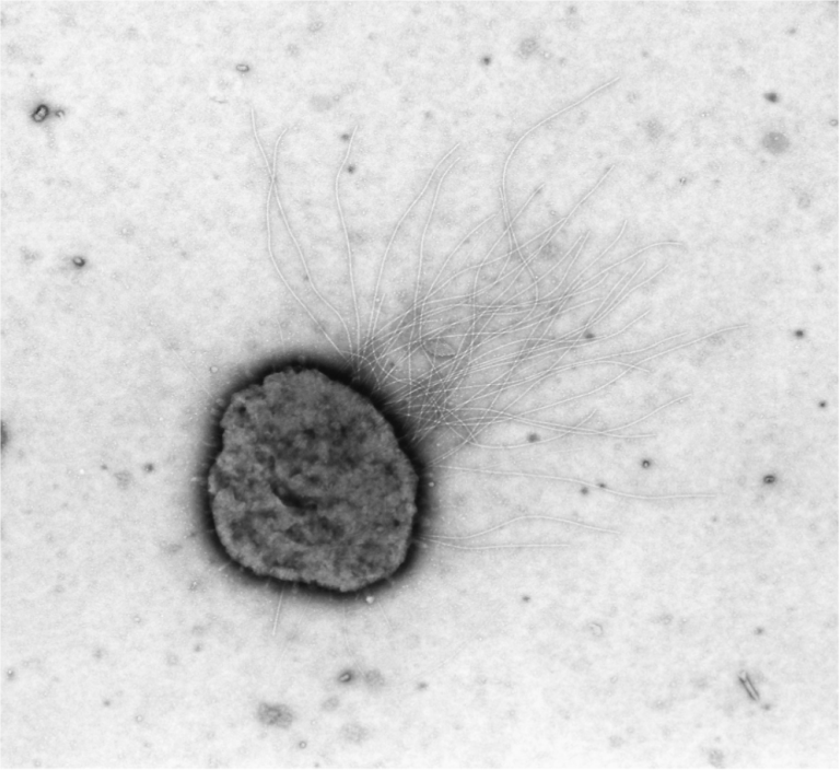
This trans­mis­sion EM of a thin sec­tion of a T2 phage in­fect­ing an E. coli cell was pro­duced by Lee Si­mon in the late 1960's. It is note­wor­thy for de­pict­ing a re­mark­ably large num­ber of prop­er­ties of the phage and of the in­fec­tion process. Can you fig­ure out how many such char­ac­ter­is­tics are il­lus­trated or at least strongly hinted at? I came up with nearly a dozen (see be­low). How about you?

I am grate­ful to Lee for let­ting me use this won­der­ful im­age.

Phage prop­er­ties il­lus­trated in this pic­ture:

  1. These phages have a head, a tail, and tail fibers.
  2. The long tail fibers are bent at a sharp an­gle.
  3. The tail is at­tached to the head by a nar­row neck col­lar.
  4. The head ap­pears to be icosa­he­dral.
  5. Ab­sorp­tion is tail first.
  6. In viri­ons ab­sorbed to the cell, the tail sheath be­comes con­tracted.
  7. In ab­sorbed viri­ons, the head ap­pears empty, al­though not al­ways to­tally.
  8. Ap­par­ently empty heads are seen in­tra­cel­lu­larly.
  9. Phages "ma­ture" in­tra­cel­lu­larly near or within the nu­cleoid.
  10. The fill­ing of the phage heads ap­pears to be a rapid process (only a few in­ter­me­di­ates be­tween empty and filled heads are seen.
  11. The host cell ap­pears to re­main in­tact through­out an ad­vanced stage of in­fec­tion (al­though small holes in the en­velopes can­not be ex­cluded).
  12. A sec­tioned in­fected E. coli looks like a pizza with pep­per­oni.


Other Posts

  • C‑Reactive Pro­tein

    Pic­tures Con­sid­ered #49 by Elio — Among the blood tests that your doc­tor may pre­scribes is the one for C‑Reactive Pro­tein (CRP). What is it? CRP is a pro­tein made by the liver in re­sponse to in­flam­ma­tion any­where in the body, so a pos­i­tive re­sult will alert the physi­cian that some­thing along those lines is go­ing on some place within you.

  • All the World Is Not E. coli

    by Elio — like most peo­ple in­ter­ested in bac­te­r­ial phys­i­ol­ogy, I spent much of my life with the nearly re­li­gious per­sua­sion that the start­ing con­di­tion for study­ing growth is when cells and their con­stituents all in­crease in uni­son. In prac­tice, such "bal­anced growth" is achieved when bac­te­ria grow ex­po­nen­tially at low con­cen­tra­tions...

  • Vi­su­al­iz­ing Cou­pled Tran­scrip­tion and Trans­la­tion in E. coli

    Pic­tures Con­sid­ered #1 by Elio — In 1970, be­fore this im­age was pub­lished, there was in­di­rect ev­i­dence that trans­la­tion in bac­te­ria is cou­pled to tran­scrip­tion – a pro­nounced de­par­ture from the eu­kary­ote way in which one process takes place in the nu­cleus while, the other is rel­e­gated to the cy­to­plasm. This clas­sic elec­tron mi­cro­graph by…

  • Sec­ondary Metabo­lites

    by Andy Bod­nar, Michelle Pri­eto, and Car­leen Vil­lar­real — The war be­tween bac­te­ria and phages has been wag­ing for eons longer than hu­mans have ex­isted. Con­se­quently, many bac­te­ria have de­vel­oped novel and unique ways for de­fend­ing them­selves against these for­eign in­vaders. Some change their sur­face pro­teins to pre­vent de­tec­tion, like chang­ing a lock on a door. Oth­ers give their own lives, ini­ti­at­ing...

  • Koch's De­vel­op­ment of Early In­sta­Gram Pos­i­tive Pho­tog­ra­phy

    Pic­tures Con­sid­ered #4 by Daniel P. Haeusser — Robert Koch is one of the key fig­ures in early bac­te­ri­ol­ogy, help­ing de­velop cul­ture tech­niques (e.g. solid me­dia), crit­i­cal rea­son­ing (e.g. Koch's pos­tu­lates), and dis­ease eti­ol­ogy (e.g.cholera and tu­ber­cu­lo­sis). He also pub­lished the first pho­tomi­cro­graphs of bac­te­ria (Fig­ure 1A) in his 1877 pa­per­Ver­fahren zur Un­ter­suchung, zum Con­servieren und Pho­tographiren…

  • Bridges Across the Periplas­mic Moat

    by Merry Youle — Gram-neg­a­tive bac­te­ria pose a par­tic­u­lar chal­lenge to any en­ter­pris­ing phage. First the phage is met by the outer mem­brane (OM) – a bar­rier to sur­mount that also can be used as a con­ve­nient hand­grip for ad­sorp­tion. Next haz­ard is the nu­cle­ase-in­fested periplasm with its jun­gle of pep­ti­do­gly­can. An in­fect­ing phage genome…

3 Comments
Oldest
Newest Most Voted
bks
12 years ago

It shows the rel­a­tive sizes of the phage and the bac­terium (which are much closer than they were in my mind's eye).
–bks

Julie Harless
12 years ago

Thank you for this!! My stu­dents are prepar­ing for an exam on dif­fer­ent types of mi­cro­scopes — how they work, how sam­ples are preped, and what can you see with each type — and I posted a link to this on the course web site. Per­fect tim­ing.
Julie Har­less
Elio replies:
How nice! I am de­lighted that you put this piece to such good use.
Elio

P. Nart
12 years ago

Won­der­ful ex­am­ple of how an im­age can be highly de­scrip­tive and ex­plana­tory of nat­ural phe­nom­ena and processes! Thanks!
Elio replies:
Noth­ing could please me more than this com­ment. This is the whole point of this se­ries, "Pic­tures Con­sid­ered!" Many thanks,
Elio