The first look at the two membranes of Gram-negatives
Pictures Considered #34
by Elio
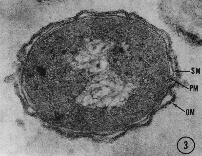
Figure 1. Three separate structures surround the cell: the outer membrane (OM), solid membrane ℠, and the plasma membrane (PM). The outer membrane, a unit membrane, appears extremely convoluted, whereas the solid membrane tightly follows the periphery of the protoplasmic constituents. Several circular structures are also evident in the outer membrane. Magnification 120,000×. Source
In a recent "Classic Spotlight" in the Journal of Bacteriology, the editor in chief, Tom Silhavy, pointed out that the first visual proof that Gram-negative bacteria have two membranes came from work by Bladen and Mergenhagen. In their 1964 paper, they clearly showed in thin section of the Gram-negative anaerobe Veilonella three envelope layers, the outer membrane, the peptidoglycan cell wall, and the cytoplasmic membrane. They went on to show that lysozyme destroys the middle layer, and, by selective lipid extraction, that lipopolysaccharide resides in the outer membrane, a term they coined. Within a few years, techniques for the separation of the inner and outer membrane by density centrifugation became available, making it possible to analyze these fractions biochemically. These confirmed the broad conclusions based on morphological studies in the original paper.